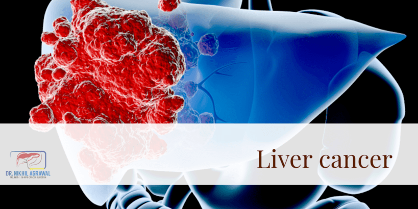
Liver cancer (Hepatocellular Carcinoma)

Primary liver cancers
A malignant (cancerous) tumour that starts in the liver is primary liver cancer. There are several types of primary liver cancers. The commonest among them is hepatocellular carcinoma. The primary liver cancers include:
Hepatocellular carcinoma: See below.
Intrahepatic bile duct cancer or cholangiocarcinoma: Read more about it here.
Angiosarcoma: Angiosarcomas are rare cancers. They begin in the cells lining the blood vessels of the liver. Risk factors for the occurrence of this cancer include chemicals (vinyl chloride, thorium dioxide, arsenic or radium) and hereditary hemochromatosis. It is aggressive cancer and usually diagnosed late, with poor survival rates.
Hepatoblastoma: Rare cancer that occurs in children. It is treated with chemotherapy and surgery.
Secondary (metastatic) liver cancers
When cancer originating from elsewhere in the body spreads to the liver, it is secondary or metastatic liver cancer. The organ where cancer originated is the primary site. Their treatment depends on their site of origin. Cancer which has metastasized to the liver portrays a poor outcome with some exceptions.
Hepatocellular carcinoma
Hepatocellular carcinoma (HCC) is a cancer of the liver cells (hepatocytes). It is the most common primary liver cancer, accounting for approximately 80% of primary liver cancers. It usually occurs in people with damaged liver (chronic liver disease and liver cirrhosis). Globally, liver cancer is the fifth most common type of cancer and the third most common cause of cancer death. It is three times more common in men.
Causes and risk factors for hepatocellular carcinoma
A risk factor is anything that increases a person's chance of developing cancer. They do not directly cause cancer, and their presence does not mean that the person will get cancer. Some people with several risk factors will never develop cancer while some without will do.
Following factors increase a person's risk of developing HCC.
- Older age: Ageing predisposes an individual to cancer.
- Gender: Hepatocellular carcinoma is more common in men.
- Cirrhosis: In cirrhosis, scar tissue replaces liver cells. There are several causes of cirrhosis. Common one includes regular alcohol intake, non-alcoholic fatty liver disease and viral hepatitis.
- Obesity, non-alcoholic fatty liver disease (NAFLD) and non-alcoholic steatohepatitis (NASH): Rising incidence of obesity worldwide has made it a major contributor to cirrhosis and liver cancer.
- Viral hepatitis: Some viruses infect the liver and cause hepatitis. They cause liver disease and also aid the development of cancer. These include hepatitis B virus (HBV) and Hepatitis C virus (HCV). HBV and HCV spread from person to person through contaminated needles, unprotected sex, and childbirth. Vaccination can prevent Hepatitis B infection. The other viruses which cause hepatitis such as hepatitis A virus and hepatitis E virus do not cause chronic hepatitis and cancer.
- Primary biliary cirrhosis: It is an autoimmune disease. It damages bile ducts in the liver, causing cirrhosis.
- Hereditary hemochromatosis: This inherited metabolic disease results in an overload of iron in the liver and cirrhosis.
- Alcohol and tobacco abuse: Alcohol causes cirrhosis. Smoking also increases the risk of liver cancer.
- Diabetes
- Toxins and Chemicals: Such as Aflatoxin, vinyl chloride and thorium dioxide (thorotrast).
- Anabolic steroids: Hormones used by some to increase their strength and muscle mass.
Signs and symptoms of liver cancer
Like other gastrointestinal cancers, hepatocellular carcinoma in early stages may not cause any symptoms.
When symptoms occur, they include:
- Pain, especially in the right upper abdomen
- Unexplained weight loss
- Reduced appetite
- Weakness or fatigue
- Jaundice (yellowing of eyes, skin and urine)
- Itching
- Nausea and vomiting
- Lump in the abdomen
- Swelling of the abdomen by the accumulation of fluid
Note that many of these symptoms can occur in diseases other than liver cancer. Having these symptoms does not mean that one has the disease. While some can harbour the disease while having none of the symptoms. If you have one or more of these symptoms, you need evaluation by a doctor, especially in the presence of risk factors.
Diagnosis (tests for liver cancer)
We use tests to diagnose and stage liver cancer. These tests include:
Clinical history and physical examination:
Blood tests: Complete blood count measures the distinct cells in the blood. Liver and kidney function tests assess the function of these organs. A liver function test may show abnormality in patients with cirrhosis. Blood clotting tests will show whether the liver is making enough of them. The blood test will also check for hepatitis B and C.
Tumour markers: A blood test will also look for a substance called Alpha-fetoprotein (AFP). 50% to 70% of patients with HCC will have high AFP. A blood test checks its level in the blood. It is a useful test to screen, diagnose and monitor.
Ultrasound: It is a basic investigation to look inside your abdomen. It uses sound waves which bounce off the internal organs and creates a picture of them on the computer monitor.
Computed tomography scan (CT scan): In this, the patient is placed in a scanner and beams of X-rays scan the abdomen from all sides. These images are then computer-processed, giving an accurate representation of inside organs. The images are enhanced with contrast by injecting it into the blood circulation. The CT scan gives information about the size and location of cancer in the liver. It shows the relation of cancer to blue duct and blood vessels in the liver. It will also detect the spread of the tumour to nearby lymph nodes and distant organs.
Magnetic resonance imaging (MRI): It is a test similar to a CT scan. Instead of X-Ray, strong magnetic fields and radio waves are used to take images. MRI is a useful modality to distinguish types of liver tumours.
Positron emission tomography (PET) scan: Cancer cells take up a larger amount of glucose. Here, injected radioactive glucose (18F-fluorodeoxyglucose; FDG) binds to the tumour, and then the patient is imaged. The images are computer-processed and combined with CT images, giving us a CT image with bright tumours.
Biopsy: Biopsy means sampling a small piece of the tumour and examining it under a microscope. Biopsy of a liver lesion is done under ultrasound or CT guidance. A biopsy is not always needed for the diagnosis of HCC. We require it only when the diagnosis is uncertain based on AFP and imaging studies.
Finding the extent of disease (Staging)
The cancer cells may break away from the primary tumour and spread through the body in one of the three ways; (a) bloodstream, (b) lymphatic system or (c) directly through the tissue.
The spread could be; local, into other parts of the liver, to the adjacent lymph nodes and the nearby organs. Or, the spread could be distant, to the lungs, bones and the peritoneum. The distant spread is metastasis.
Staging is a systematic way to describe the extent of disease. It tells clinicians about the local and distant spread of disease and helps them plan the appropriate treatment. It also predicts the outcome of treatment, a lower stage is associated with a better outcome. Liver cancer is staged based on the tests described in the previous section.
Several staging systems are used for liver cancer staging.
Tumour node and metastasis (TNM) staging system by American joint committee on Cancer (AJCC)
Developed by the American Joint Committee on Cancer (AJCC), it is used for classification of the stage. It is based on the following three key elements and span from I to IV.
The extent (size) of the tumour (T): What is the size of the tumour? How many tumours are there? Is it invading nearby vessels? Has cancer reached nearby structures or organs?
The spread to nearby lymph nodes (N): Has cancer spread to nearby lymph nodes? And to how many?
The spread (metastasis) to distant sites (M): Has cancer spread to distant lymph nodes or distant organs such as the peritoneum, bones or lungs?
Numbers and letters after T, N and M give further details. Higher the number, more advanced the tumour. Combined Information from T, N and M assigns an overall stage, a process called stage grouping. Liver cancer stage ranges from I to IV. Stages I to III are localized disease and stage IV is advanced cancer (metastatic disease). Chances of recovery from cancer (prognosis) depend on the stage of the disease at the time of diagnosis. Lower the stage, better is the long-term prognosis.
Barcelona clinic liver cancer system (BCLC)
The BCLC system stratifies HCC patients based on tumour size, extent, liver function, and performance status. BCLC stage grouping includes very early stage (stage 0), early stage (stage A), intermediate stage (stage B), advanced stage (stage C) and terminal stage (stage D).
The Cancer of the Liver Italian Program (CLIP) system
The Okuda system
Besides the stage, the function of the liver is an important consideration in managing these patients.
The staging system described above gives us an accurate representation of the disease burden and likely outcome.
Practically, we can classify them into a few broad categories depending on whether they can be surgically removed.
- Resectable or transplantable
- Locally advanced or unresectable
- Metastatic
Treatment
In cancer care, doctors from different specialities work together to plan patients' overall treatment. This is called a multidisciplinary team. This ensures the best outcome for the patient. The treatment of liver cancer depends on the condition of the liver and the stage of cancer. The patient's overall health is also an important consideration.
In early tumours with a good functioning liver, treatment aims at eliminating cancer and curing the patient. In advanced stages with poorly functioning liver, the aim of treatment changes to slowing growth of the tumour, relieving symptoms and improving quality of life.
Treatment to eliminate and potentially cure
Surgery for liver cancer
Surgery for liver cancer involves the removal of cancer. It is the most effective treatment option, if feasible. It can only be applied when the tumour has not spread outside the liver. Two types of surgical procedures are used to treat liver cancer.
Hepatectomy or liver resection
Hepatectomy is the removal of part of the liver containing cancer with healthy margins. It is done when the remaining liver after surgery is of adequate size and functioning well. The remaining liver grows over a few weeks. With advancements in surgical technique, we do this procedure laparoscopically in selected patients.
Open surgery uses a long incision over the abdomen for the surgery. The laparoscopic approach uses minimally invasive techniques to do the same surgery with tiny incisions. This entails the insertion of special long thin surgical tools through these small holes. Laparoscopic liver resection results in faster recovery and reduced pain compared to conventional open surgery.
Liver transplantation
Majority of hepatocellular carcinoma occur in people with cirrhosis. Many liver cancers in these patients are not amenable to resection because the remaining liver is not of adequate size or not functioning properly. Liver transplant is an option in these patients. However, not every patient is a candidate for a transplant. There are strict criteria which include tumour size and number.
Ablation
Ablation uses extreme heat, cold or chemical to kill the tumour cells. These can be used in patients with small tumours or when surgery is not a good option because of poor liver function, or poor health of the patient, or location of the tumour in the liver. This is best for small tumours which are less than 2 cm. But it can be used in tumours up to 5 cm, though it is less effective. To improve efficacy in larger tumours, it can be combined with embolization techniques. Radiofrequency ablation (RFA) uses high-frequency radio waves to generate heat and kill the tumour. A probe is inserted into the tumour guided by ultrasound or CT scan. Microwave ablation uses microwaves to generate heat and kill the tumour. Cryoablation or cryotherapy kills the tumour by freezing it with a metal probe. Percutaneous ethanol injection (PEI) can also be given into the tumour killing the cells.
Radiation therapy
Radiation therapy uses high energy x-rays to kill tumour cells. Stereotactic body radiation therapy (SBRT) is a technique to deliver high doses of radiation to the tumour while sparing nearby tissues and organs. Radiation therapy is used in tumours which are not amenable to surgery.
Treatment to palliate
These treatment options are chosen when the disease is not amenable to curative treatment. Sometimes, they are instituted as a temporary measure, while the patient is optimized for surgery.
Embolization therapy
Trans-arterial chemoembolization (TACE) or trans-arterial radioembolization (TARE)
Embolization means blocking the blood supply of the tumour. A catheter is passed into the vessel supplying the tumour and is blocked with small particles and other agents. Chemotherapeutic agents and radioactive beads can also be delivered directly into the tumour during this, called chemoembolization or radioembolization. Embolization is an option for patients whose tumours cannot undergo operation or ablation.
Targeted therapy
Substances which identify and attack cancer cells without harming normal cells. It targets cancer’s specific genes, proteins, or the tissue environment responsible for cancer growth. Tests are required to identify the targets.
For HCC, anti-angiogenesis drugs are most commonly used. These retard the formation of new blood vessels, which delivers nutrients for the growth of tumours and thus stops the cancers from growing.
Immunotherapy
It uses the patient's immune system to fight cancer. Immune checkpoint inhibitor therapy is a type of immunotherapy. These block the pathways through which the cancer cells hide from the body's immune system.
Prognosis
Survival rates
The 5-year survival rate is the percent of people who are alive 5 years after the diagnosis of cancer. The general 5-year survival rate of hepatocellular carcinoma is 18%. For localized cancers, the 5-year survival is 40-50% after treatment.
Screening for liver cancer
Screening for cancer means looking for cancer in apparently healthy individuals. The aim is to detect cancer before someone develops signs and symptoms of it. If we can detect cancer earlier, we can treat it better.
Screening for liver cancer is done in patients who have diseased liver such as cirrhosis and viral hepatitis. Screening options include testing blood for alpha fetoprotein (AFP) and imaging studies like an ultrasound, CT scan or MRI.
How to lower your risk of HCC
We can decrease the risk of developing liver cancer. A vaccine can protect against Hepatitis B. Cirrhosis can be avoided by decreasing the consumption of alcohol and maintaining a healthy weight. Fatty liver and associated NAFLD can be prevented by regular exercise, eating a good diet and maintaining a healthy weight. Treatment of hepatitis B and C is now available. Adequate treatment of hepatitis can also help reduce the risk of developing HCC.
- Don’t indulge in smoking and drinking
- Get vaccinated for hepatitis B
- If you have hepatitis, get treated
- Maintain a healthy weight (BMI of 23-25)
- Regularly get checked (screening) for liver cancer if you are at high risk
Wish you a speedy recovery!

