Liver (hepatic) cyst
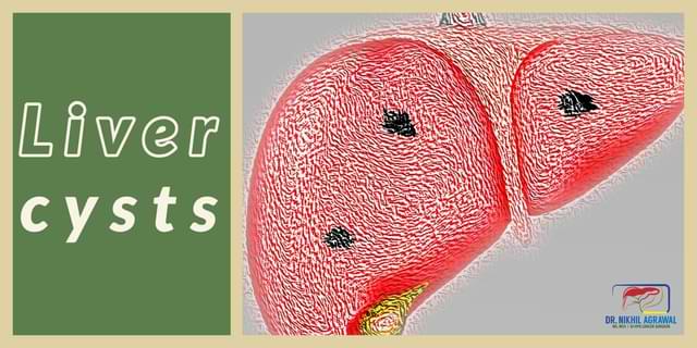
A cyst is a thin-walled sac filled with fluid or semisolid material. Many types of cysts can form in the liver.


The liver
The liver is a large reddish-brown organ. It is in the right upper abdomen underneath the rib cage. It has two sections, the right and left lobe. The liver is essential for digestion, getting rid of toxins from the body and synthesizing proteins and biochemicals. It produces bile which is collected by bile ducts and transported to the intestine. The liver is made of cells called hepatocytes. The bile ducts and blood vessels criss-cross the entire liver.
Many types of tumours and cysts can form in the liver. The tumours can be benign (noncancerous) or cancerous (malignant).
Liver (hepatic) cysts
A cyst is a thin-walled sac filled with fluid or semisolid material. With the increasing use of imaging modalities like ultrasound, CT and MRI, liver cysts are now frequently detected.
Many types of cysts can form in the liver. These include:
- Simple cysts
- Polycystic liver disease
- Parasitic or hydatid (echinococcal cysts)
- Cystic tumours
- Liver abscesses
- Choledochal cysts and Caroli disease
Liver cysts are found in approximately 5% of the people. Only 10-15% of them will be symptomatic. Majority of these cysts are benign (noncancerous). They can be congenital (present since birth) or can develop later.
They can be found incidentally when tests done for a routine check-up or testing other diseases picks it up. They can also cause symptoms and are then found in further testing.
Simple hepatic cysts
Simple hepatic cysts are common. They are congenital and result from the malformation of bile ducts. They are typically single but can also be multiple. They are often asymptomatic and do not require any treatment. Rarely they can cause symptoms and can have complications. These include compression of blood vessels, bile ducts, or adjacent organs. Bleeding or infection can occur in these. They can rupture or turn cancerous.
These cysts do not require any treatment except in rare cases when they are symptomatic or complicated.
Polycystic liver disease (PCLD)
It is a rare inherited condition. In PCLD cysts form throughout the liver. The cysts can occur only in the liver or be associated with cysts in the kidney; polycystic kidney disease. The cysts can be in small or large grape-like clusters, or they can grow independently in other parts of the liver. It can rarely progress to hepatic fibrosis, portal hypertension, and liver failure.
If the cysts are not very large and not causing symptoms, they are observed. The indication for surgery depends on symptoms and complications.
Hydatid cyst
Hydatid cyst is a parasitic cyst of the liver. It is formed when ingested eggs of a parasite Echinococcus granulosus (animal tapeworm) develop into larvae, reach the human liver and then become encysted and grow. The tapeworm is found in the intestines of cattle, sheep and dogs.
These cysts continue to grow and have a tendency to complicate. They can get infected, can rupture into the bile duct and cause their blockage, or can rupture in your abdomen or chest.
These cysts need treatment. The treatment involves removal of cyst content along with instillation of chemicals (sclerosants) to destroy the parasitic lining of the cyst. It can be achieved surgically or through percutaneous radiological techniques. The commonly used radiological procedure is PAIR (percutaneous puncture, aspiration, injection, reaspiration). The surgical procedure is laparoscopic partial cystectomy, enucleation, pericystectomy or liver resection. These procedures are now done laparoscopically.
Liver abscesses
Liver abscess is pus-filled cyst inside the liver. It can be amoebic or bacterial. Those having liver abscess present with pain, fever and increased leukocyte counts. Liver abscesses can cause a generalised infection called sepsis and can also rupture. They are treated with antibiotics and/or antiamoebics. They may also require percutaneous aspiration or tube placement into the abscess to drain the pus.
Cystic tumours (biliary cystadenoma and cystadenocarcinoma)
Some primary or metastatic liver tumours with central necrosis can appear as liver cysts. Cystic tumour of the liver is biliary cystadenoma and cystadenocarcinoma. It is an uncommon tumour. It is usually large, multiloculated and has internal septations. These cysts more commonly occur in middle age women. Hepatic cystadenoma can be serous or mucinous. They can be symptomatic and the symptoms include abdominal pain and/or discomfort, with distension and a palpable mass. They can also complicate with bleeding, rupture and compression of adjacent structures.
These cysts are premalignant and can transform into cancer (cystadenocarcinoma). Complete resection is the treatment of choice.
Signs and symptoms of liver cysts
The liver cysts are mostly asymptomatic. Symptoms are rarely present, but when present they include:
- Abdominal pain
- Abdominal discomfort
- Feeling of fullness
- Abdominal bloating
- Nausea or vomiting
- Loss of appetite
- Early satiety
- Mass in the abdomen
- Jaundice caused by bile duct obstruction
- Ascites (water in the abdomen)
- Unexplained weight loss
Diagnosis / Workup for liver cyst
Treatment of these liver cysts depends on symptoms, size, and type of lesion. It is important to identify the type of cyst. Tests help to diagnose and differentiate one cyst from another. Tests are also required to monitor them if we choose to keep them under observation.
History and physical examination: Understanding symptoms and checking for signs by a physician are basics of arriving at a diagnosis.
Blood tests: Complete blood count measures the distinct cells in the blood. Liver and kidney function tests assess the function of these organs. A liver function test is usually normal. Blood clotting tests will show whether the liver is making enough of them. The blood test will also check for hepatitis B and C. Hydatid cyst patients may have elevated eosinophil counts and echinococcal antibody titres. Amoebic liver abscess patients will have antibodies against Entamoeba histolytica.
Tumour markers: A blood test will also look for tumour markers. Tumour markers are elevated in malignant tumours and help differentiate benign tumours from malignant.
Ultrasound: It is a basic investigation to look inside your abdomen and usually the first investigation which detects the cyst in the liver. It uses sound waves which bounce off the internal organs and creates a picture of them on the computer monitor.
Computed tomography scan (CT scan): In this, the patient is placed in a scanner and beams of X-rays scan the abdomen from all sides. These images are then computer-processed, giving an accurate representation of inside organs. For liver lesions, triple-phase CT helps us determine the type of lesion. In this, the images are enhanced with contrast by injecting it into the blood circulation and scanning is done in phases while the contrast passes through the liver. The CT scan gives information about the size and location of cysts in the liver. It also shows the relation of cysts to blue duct and blood vessels in the liver.
Magnetic resonance imaging (MRI): It is a test similar to a CT scan. Instead of X-Ray, strong magnetic fields and radio waves are used to take images. MRI is a useful modality to distinguish types of liver cysts. MR images are also enhanced with liver-specific contrast agents, acquiring images in phases.
Biopsy: Biopsy means sampling a small piece of the tumour and examining it under a microscope. Biopsy of a liver lesion is done under ultrasound or CT guidance. A biopsy is not usually needed for the diagnosis of liver cysts.
Cyst fluid aspiration: Cyst fluid can be aspirated and tested. However, it is generally avoided.
Treatment of liver cysts
Treatment of these cysts depends on the type of cyst, their size and the presence or absence of symptoms. Hydatid cyst, biliary cystadenoma, cystadenocarcinoma and liver abscesses always need treatment. While other cysts can be kept under observation. It is essential to establish an accurate diagnosis, determine the nature of lesions and symptoms of the patient before formulating a management plan.
When treatment is required, the treatment options include:
- Medicines
- Percutaneous image (ultrasound or CT) guided treatment
- Surgical treatment
Medicines
Antibiotics and antiamoebic agents are used to treat a liver abscess. Antihelminthics are used for hydatid cyst. Antihelminthics alone are effective alone only in the early stage of the hydatid disease. They are typically used in the perioperative period, along with surgery or percutaneous treatment.
Percutaneous image (ultrasound or CT) guided treatment
Some of these cystic lesions can be treated with image-guided interventions. Hydatid cyst can be treated with a procedure called PAIR (puncture, aspiration, injection, reaspiration). The liver abscess can be treated with percutaneous needle aspiration or pigtail catheter drainage. Some simple cysts can be treated with aspiration combined with sclerosis but can have a high recurrence and failure rates.
Surgical treatment
Some of these cysts will require surgical intervention.
Surgery for liver cysts
Simple cyst and PCLD
Simple liver cyst or the cysts of PCLD requires treatment when they are causing symptoms. The surgery for these cysts involves cutting out the part of the cyst which is on the surface of the liver, called fenestration or unroofing.
Neoplastic cysts
For neoplastic cysts such as cystadenoma and adenocarcinoma, the surgery entails complete excision of the cyst. This could be done by enucleation in which only the cyst is removed from the liver or liver resection in which the part of the liver containing the cyst is removed.
Hydatid cyst
In the surgical treatment of hydatid cyst, the cyst contents are removed, and the parasite is killed with scolicidal agents such as hypertonic saline, chlorhexidine, absolute alcohol, and cetrimide. The cyst is then partially excised; partial cystectomy. Some lesions will require a different approach because of their anatomical location or complication. These include pericystectomy, enucleation and liver resection.
Laparoscopic or robotic surgery for the liver cyst
Many of the surgical procedures mentioned above are now done laparoscopically or robotically. These approaches are minimally invasive. Here the surgery is done through small holes instead of large incisions. This gives quicker recovery with lesser pain.
Freqently asked questions

About Author
Dr. Nikhil Agrawal
MS, MCh
Dr. Nikhil Agrawal is a leading GI-HPB Surgical Oncologist with 20+ years of experience in complex cancers of the esophagus, stomach, colon, rectum, liver, pancreas, gallbladder, and bile ducts. He leads the GI-HPB Oncology Program at Apollo Hospitals, Delhi and Gurugram, with expertise in advanced robotic and laparoscopic cancer surgery.
His practice focuses on evidence-based, multidisciplinary care with an emphasis on individualized treatment and long-term outcomes.
He trained at BHU, SGPGI Lucknow, AIIMS New Delhi, and SNUBH, South Korea, and is a robotic surgery proctor who trains surgeons in advanced GI-HPB cancer surgery. He is also regularly invited as faculty at national and international scientific meetings.
This website helps patients and families understand GI and HPB diseases and cancers, treatment options, and what to expect during recovery and long-term care.